Do kidney stones and CKD need different care?
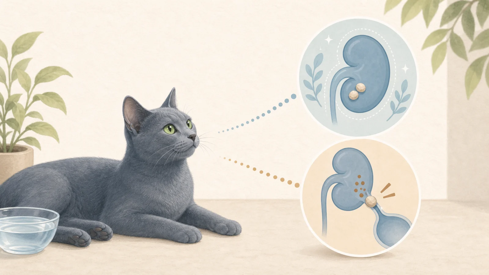
On the way out of a routine ultrasound, the chart says: "Nephroliths (kidney stones) noted." Two questions arrive at once. Will this make the kidney disease go faster? Do I have to change everything I have been doing?
The answer depends on where the stone is. The same stone sitting quietly inside the kidney leaves your CKD care almost untouched; the same stone lodged in the ureter (the thin tube from kidney to bladder) is a different disease altogether. This article lays out that fork in the road.
1. Where is the stone — in the kidney, or in the ureter?
More than 90% of upper urinary tract stones in cats are calcium oxalate (ACVIM 2016). That single fact runs through this whole article: calcium oxalate cannot be dissolved by diet or medication. The story of a bladder struvite stone that "melted away on a dissolution diet" does not apply here.
| Nephrolith (inside the kidney) | Ureterolith (blocking the ureter) | |
|---|---|---|
| Location | Renal pelvis (where urine collects) or parenchyma | Inside the tube running down to the bladder |
| Signs | Usually none — found incidentally on imaging | Sudden vomiting, loss of appetite, pain, lethargy, values jumping |
| Effect on CKD | No significant difference in progression or survival (Ross 2007) | The blocked kidney loses function within days to weeks |
| Urgency | Low — monitor | Emergency — time is kidney tissue |
| Response | Treat only if it causes a problem | 24–72 h medical trial → SUB or stent if it does not pass |
2. Stones inside the kidney — the rule is to leave them alone
First, how common this is. In a Japanese referral hospital, 73% of 140 CKD cats had kidney or ureteral stones (Hsu 2022 — a referral population, so probably an overestimate). In a UK first-opinion cohort, 61% of CKD cats showed nephrocalcinosis (mineral deposits in kidney tissue) on ultrasound, rising to 81% in cats with hypercalcaemia (Tang 2024). In other words, "stones seen" is a common companion finding in CKD, not an exception.
So do these stones speed up the kidney disease? One study asked exactly that. CKD cats with and without nephroliths were followed for two years (Ross 2007): the rate of creatinine rise, death from renal causes, and overall survival were not significantly different between the groups. On that basis the ACVIM consensus states:
So with a nephrolith, the CKD care you are already doing — phosphorus, hydration, blood pressure, body weight — does not change. What changes is the list of things you watch, covered below.
3. Stones in the ureter — the disease that quietly takes one side
The trouble starts when a stone drops from the kidney into the ureter and lodges there. A cat's ureter has an internal diameter of roughly 1 mm, so a 2–3 mm stone is plenty. And here is the trap every CKD caregiver needs to know.
A single blocked ureter usually causes no signs. The other kidney picks up the work, so bloodwork barely moves. The blocked kidney quietly shrinks over weeks while the other enlarges — the picture radiologists call "big kidney–little kidney". A cat found in this state has already lost one kidney, and if a stone later lodges on the remaining big side, that becomes bilateral obstruction: no urine leaving the body, a true emergency.
· Was eating yesterday, now refuses food and vomits
· Resents having the belly touched, hunches, hides
· Urine volume drops, or there is blood in the urine
· Recheck shows creatinine far above last month's (a jump, not the gentle CKD slope)
CKD moves in units of months. When things worsen in units of days, suspect a blockage first.
On ultrasound the key measurement is the width of the renal pelvis (the urine-collecting space inside the kidney). Normal cats, and cats on fluids, can show up to about 3.5 mm; 3.5 mm or more warrants further investigation, and 13 mm or more almost always means obstruction (Griffin 2020). Recent work has also described obstructed cats with barely dilated pelves, so clinicians judge on the combination — "sudden decline + visible ureter + one kidney enlarged" — rather than one number.
4. When it is blocked — the medical trial, and the SUB
A blocked ureter does not mean straight to surgery. The ACVIM consensus allows a 24–72 hour medical trial first: fluids to increase urine flow, drugs that relax the ureter (prazosin and similar), diuretics and analgesia as needed — waiting for the stone to pass into the bladder on its own.
Expectations should be realistic, though. In a 2023 study of 72 cats (103 kidneys) treated medically only, stone obstructions resolved in 23% (Merindol 2023). The successes shared features:
- Small stones — median 1.85 mm in successes vs 3 mm in failures
- Distal position (near the bladder) — roughly double the success rate
- Younger cats with still-large kidneys — that is, kidneys with reserve left
Put the other way: for an older CKD cat with a stone over 3 mm lodged high up, days spent on a medical trial can simply be days of kidney tissue lost. That is why the consensus states that obstructed ureters should be relieved (stent or bypass) to restore function, and that because interventional procedures carry lower morbidity and mortality than traditional surgery, referral is advised.
| Option | What it does | Worth knowing |
|---|---|---|
| Medical management | Fluids, ureteral relaxants, analgesia | 24–72 h limit. Only about one in four stone obstructions pass |
| Ureteral stent | A fine tube placed inside the ureter | Feline ureters are so narrow that irritation and re-obstruction are common; largely superseded by the SUB |
| SUB (subcutaneous ureteral bypass) | A new route kidney → port under the skin → bladder, bypassing the ureter | The standard procedure for feline ureteral obstruction. Needs port flushing every 3 months for life |
| Ureterotomy | Opening the ureter to remove the stone | Higher leakage and re-obstruction risk; no longer first choice |
Placing a SUB is the beginning of management, not the end. The two long-term complications are mineralisation (deposits inside the device) and chronic urinary tract infection. In a 2025 follow-up of 66 cats and 95 devices, cats flushed with saline alone had 32% mineralisation and 33% chronic infection, whereas those flushed with 2% tEDTA solution had 19% and 3% respectively (Berent 2025). If your cat has a SUB, it is worth asking what the flushing solution is.
5. So what actually changes in the care?
This is the heart of the article. Care for a CKD cat with stones is "CKD care + calcium oxalate prevention" — and the two point the same way almost everywhere, colliding in exactly one place.
| Item | CKD alone | CKD with stones |
|---|---|---|
| Water | Important | More important — dilute urine is the only real "preventive". Wet food, fountains, subcutaneous fluids if needed |
| Therapeutic diet | Renal diet | Stay on the renal diet. Its phosphorus restriction and alkalinising tendency also suit oxalate prevention. But "urinary" diets designed for struvite (acidifying) are contraindicated |
| Urine pH | No specific target | Avoid drifting acidic — target 6.6–7.5. No acidifiers, no cranberry-type supplements |
| Calcium | Often only total calcium checked | Measure ionised calcium (iCa) — the real cause of the stones may be hiding here |
| Supplements | Phosphate binders, omega-3 etc. | Re-examine calcium-based binders, vitamin D, high-dose vitamin C. Potassium citrate may help (vet's call) |
| Imaging | As needed | Scheduled ultrasound — track stone position, size, pelvic width |
| Sudden decline | Recheck, fluids | Ultrasound first — rule out obstruction |
In short, what the cat eats barely changes; what you look at expands. There is no reason to stop a renal diet because of stones — switching to an acidifying "urinary" diet is, if anything, the way to grow calcium oxalate.
6. The one collision — phosphorus restriction and calcium
Renal diets lower phosphorus. Lower phosphorus raises the relative calcium ratio (Ca:P), and in some cats ionised calcium climbs above normal — hypercalcaemia. In the Tang 2024 study the strongest independent risk factor for nephrocalcinosis was precisely ionised calcium, and in Hsu 2022 the CKD cats with stones had the same total calcium but higher ionised calcium and higher urinary calcium excretion.
What if ionised calcium is high? Fortunately the first tool is not a drug but diet adjustment. A 2024 study reported ionised calcium normalising with dietary change alone in cats with idiopathic hypercalcaemia or CKD (Ehrlich 2024). The direction is "keep phosphorus low but bring calcium down too" — the targets proposed for hypercalcaemic cats are calcium below 2,000 mg/1000 kcal and Ca:P below 1.4. Several therapeutic diets meet both, and the choice is made with your vet, with the cat's phosphorus and calcium numbers side by side.
7. Monitoring — what, and how often
- Ultrasound — for a silent nephrolith, every 6 months as a baseline; tighten to 3 months if the stone is growing or the pelvis is widening. Record three things: position (pelvis/ureter), size (mm), pelvic width (mm). How those three numbers move over time is the whole story
- Ionised calcium — once when stones are found, then whenever the renal diet changes or a calcium-containing supplement is started
- Urinalysis — specific gravity (is it staying dilute?), pH (drifting acidic?), and infection. Stones shelter bacteria; infection in turn grows stones. If pyelonephritis is suspected, culture the urine
- The slope of creatinine — watch the change since the last test more than the value itself. A cat that used to rise 0.2 at a time and suddenly jumps 1.0 is not progressing; something has happened
8. What to ask your vet
- "Is the stone inside the kidney or in the ureter? How many mm, and what is the pelvic width?"
- "Is this stone blocking urine flow, or sitting where it could? Or is it in a place we can watch?"
- "Have we ever measured ionised calcium? Not total calcium."
- "Can we stay on the renal diet? You are not suggesting a urinary diet, are you?"
- "Some of the binders and supplements we give contain calcium or vitamin D — is that all right?"
- "When is the next ultrasound? And which signs before then mean come in straight away?"
- "If it does obstruct, which hospital places SUBs? May I find out in advance?"
Considering magnesium during calcium follow-up
Magnesium (Mg) is another part of mineral assessment during renal feeding. A 2024 randomised feline trial evaluated calcium and FGF23 during a magnesium-enriched, phosphorus-restricted diet in CKD cats without hypercalcaemia or hypermagnesaemia. It assessed mineral-marker changes; stone recurrence and longer-term outcomes require further evaluation. Feline clinical trial
To explore this approach, bring Mg, iCa and phosphorus results together with food and supplement records. Agree an appropriate dietary choice and recheck plan; monitor blood Mg if supplementation is selected. Record proportions of foods eaten, intake and change dates to compare subsequent calcium results. Check whether an existing report already includes Mg.
9. Related
- Stages and target values — the baseline that holds regardless of stones
- Uraemic symptoms — where "progression" and "obstruction" overlap and where they part
- Subcutaneous fluids at home — why water matters even more with stones
- Getting the most from your vet — how to communicate a sudden change